Pal cadaver axial skeleton skull lab practical question 25 presents a unique opportunity to delve into the intricate anatomy of the human skull. This comprehensive guide will provide a detailed overview of the question, offering a step-by-step approach to answering it accurately.
Furthermore, it will explore the comparative anatomy of the skull, highlighting similarities and differences across species, and discuss the clinical applications of skull anatomy in various fields.
The human skull is a complex and fascinating structure composed of multiple bones that work together to protect the brain and other vital organs. Understanding the anatomy of the skull is crucial for medical professionals, forensic scientists, and anthropologists. This guide will provide a comprehensive overview of the bones that make up the skull, their functions, and the methods used to examine them in a pal cadaver lab setting.
Anatomical Structures of the Axial Skeleton Skull: Pal Cadaver Axial Skeleton Skull Lab Practical Question 25

The skull, a complex bony structure, serves as a protective enclosure for the brain and other vital organs. Composed of 22 bones, the skull can be divided into two main regions: the cranium and the facial skeleton.
The cranium, forming the upper portion of the skull, comprises eight bones: the frontal bone, two parietal bones, two temporal bones, the occipital bone, the sphenoid bone, and the ethmoid bone. These bones interlock to form a protective vault around the brain.
The facial skeleton, located anteriorly, consists of 14 bones that contribute to the formation of the face. These bones include the two nasal bones, two maxillae, two zygomatic bones, two lacrimal bones, two palatine bones, two inferior nasal conchae, the vomer, and the mandible.
Each bone of the skull plays specific roles in protecting and supporting the underlying structures. The frontal bone forms the forehead and contributes to the roof of the eye sockets, while the parietal bones form the majority of the skull’s calvaria.
The temporal bones house the organs of hearing and balance, and the occipital bone forms the posterior aspect of the skull and provides attachment for muscles.
The sphenoid bone, located at the base of the skull, contributes to the formation of the middle cranial fossa and provides passageways for nerves and blood vessels. The ethmoid bone, also located at the base of the skull, forms part of the nasal cavity and contributes to the formation of the orbits.
The facial skeleton provides support for the facial muscles and sensory organs. The maxillae form the upper jaw and contribute to the floor of the orbits, while the zygomatic bones form the cheekbones and provide attachment for muscles of facial expression.
The nasal bones form the bridge of the nose, and the lacrimal bones form part of the medial wall of the orbits. The palatine bones contribute to the formation of the hard palate, and the inferior nasal conchae are scroll-like bones that contribute to the formation of the nasal cavity.
The vomer forms part of the nasal septum, and the mandible forms the lower jaw and provides attachment for muscles of mastication.
Pal Cadaver Axial Skeleton Skull Lab Practical Question 25
Question: Identify the following structures on the pal cadaver skull: frontal bone, parietal bone, temporal bone, occipital bone, sphenoid bone, ethmoid bone, nasal bone, maxilla, zygomatic bone, lacrimal bone, palatine bone, inferior nasal concha, vomer, and mandible.
Approach:
- Familiarize yourself with the anatomical structures of the skull.
- Locate the frontal bone, which forms the forehead and the roof of the eye sockets.
- Identify the parietal bones, which form the majority of the skull’s calvaria.
- Locate the temporal bones, which house the organs of hearing and balance.
- Identify the occipital bone, which forms the posterior aspect of the skull.
- Locate the sphenoid bone, which is located at the base of the skull and contributes to the formation of the middle cranial fossa.
- Identify the ethmoid bone, which is also located at the base of the skull and contributes to the formation of the nasal cavity and the orbits.
- Locate the nasal bones, which form the bridge of the nose.
- Identify the maxillae, which form the upper jaw and contribute to the floor of the orbits.
- Locate the zygomatic bones, which form the cheekbones.
- Identify the lacrimal bones, which form part of the medial wall of the orbits.
- Locate the palatine bones, which contribute to the formation of the hard palate.
- Identify the inferior nasal conchae, which are scroll-like bones that contribute to the formation of the nasal cavity.
- Locate the vomer, which forms part of the nasal septum.
- Identify the mandible, which forms the lower jaw.
Methods and Procedures for Pal Cadaver Skull Examination
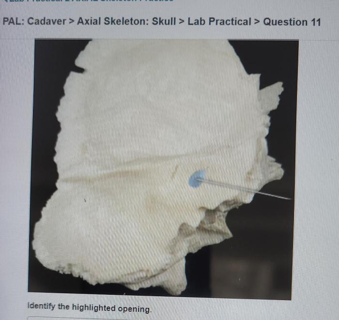
Table of Methods and Procedures:
| Method/Procedure | Description | |—|—| | Visual inspection | Examination of the skull using the naked eye and a magnifying glass to observe its shape, size, and any abnormalities. | | Palpation | Examination of the skull using the fingers to feel its texture, contours, and any irregularities.
| | Percussion | Examination of the skull using gentle tapping to assess its density and thickness. | | Auscultation | Examination of the skull using a stethoscope to listen for any unusual sounds, such as crepitus or bruit. | | Radiography | Examination of the skull using X-rays to visualize its internal structures and identify any fractures or other abnormalities.
| | Computed tomography (CT) | Examination of the skull using a series of X-rays taken from different angles to create cross-sectional images of its internal structures. | | Magnetic resonance imaging (MRI) | Examination of the skull using a strong magnetic field and radio waves to create detailed images of its soft tissues and structures.
|
Essential Tools and Materials:
– Pal cadaver skull – Gloves – Magnifying glass – Stethoscope – X-ray machine – CT scanner – MRI scanner
Importance of Proper Handling and Preservation Techniques:
Proper handling and preservation techniques are essential to maintain the integrity of the pal cadaver skull and ensure accurate examination. The skull should be handled with care to avoid damage or contamination. It should be stored in a cool, dry place and protected from moisture and pests.
Comparative Anatomy of the Skull

The anatomy of the human skull exhibits similarities and differences compared to the skulls of other mammals. The basic structure of the skull, including the presence of a cranium and a facial skeleton, is conserved across mammalian species.
Similarities:
– The cranium of all mammals is composed of eight bones: the frontal bone, two parietal bones, two temporal bones, the occipital bone, the sphenoid bone, and the ethmoid bone. – The facial skeleton of all mammals consists of 14 bones, including the two nasal bones, two maxillae, two zygomatic bones, two lacrimal bones, two palatine bones, two inferior nasal conchae, the vomer, and the mandible.
– The skull provides protection for the brain and other vital organs, and supports the facial muscles and sensory organs.
Differences:
– The size and shape of the skull varies significantly among mammalian species, reflecting adaptations to different ecological niches and lifestyles. – The development of specialized structures, such as antlers or horns, can modify the appearance of the skull. – The dentition of mammals varies widely, and the structure of the skull reflects these differences in feeding habits.
Evolutionary Significance:
The comparative anatomy of the skull provides insights into the evolutionary relationships and adaptations of different mammalian species. Similarities in skull structure suggest common ancestry, while differences reflect adaptations to specific environments and lifestyles.
Clinical Applications of Skull Anatomy
Knowledge of skull anatomy is essential in various clinical fields, including neurosurgery and forensic science.
Neurosurgery:
– Skull anatomy guides neurosurgeons in performing delicate procedures on the brain and its surrounding structures. – Understanding the location and relationships of skull bones and foramina is crucial for safe access to the brain. – Skull anatomy also aids in the diagnosis and treatment of skull fractures and other traumatic injuries.
Forensic Science:
– Skull anatomy assists forensic anthropologists in identifying human remains and estimating the age, sex, and ancestry of individuals. – Analysis of skull features, such as the shape of the orbits and the presence of certain anatomical landmarks, can provide valuable information for identification purposes.
– Skull anatomy is also used in facial reconstruction, a technique employed to restore the facial features of individuals whose remains are incomplete or damaged.
Questions Often Asked
What is the purpose of the foramen magnum?
The foramen magnum is a large opening at the base of the skull that allows the spinal cord to connect to the brain.
What are the three main regions of the skull?
The three main regions of the skull are the cranium, the face, and the mandible.
What is the difference between a suture and a fontanelle?
A suture is a fibrous joint that connects the bones of the skull, while a fontanelle is a soft spot where the bones of the skull have not yet fused together.