Label the veins of the head and trunk – Labeling the veins of the head and trunk is an essential aspect of understanding the human circulatory system. This comprehensive guide provides a detailed overview of the veins of the head and trunk, including their location, tributaries, and clinical significance.
The veins of the head and trunk play a crucial role in returning deoxygenated blood to the heart. Understanding their anatomy is vital for medical professionals, students, and anyone interested in the human body.
Head Veins
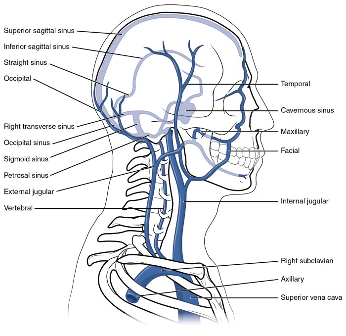
The veins of the head can be divided into two groups: those that drain the scalp and those that drain the face. The veins that drain the scalp are the superficial temporal vein and the posterior auricular vein. The superficial temporal vein drains the lateral aspect of the scalp, while the posterior auricular vein drains the posterior aspect of the scalp.
The veins that drain the face are the facial vein and the internal jugular vein. The facial vein drains the anterior aspect of the face, while the internal jugular vein drains the posterior aspect of the face.
External Jugular Vein
The external jugular vein is a large vein that drains the lateral and anterior aspects of the face and neck. It begins at the angle of the mandible and runs down the neck, just lateral to the sternocleidomastoid muscle. The external jugular vein receives tributaries from the facial vein, the retromandibular vein, and the occipital vein.
It terminates by joining the subclavian vein.
Internal Jugular Vein
The internal jugular vein is a large vein that drains the posterior aspect of the face and neck. It begins at the jugular foramen and runs down the neck, deep to the sternocleidomastoid muscle. The internal jugular vein receives tributaries from the facial vein, the lingual vein, the pharyngeal vein, and the occipital vein.
It terminates by joining the subclavian vein.
Facial Vein
The facial vein is a large vein that drains the anterior aspect of the face. It begins at the medial canthus of the eye and runs down the face, just lateral to the nose. The facial vein receives tributaries from the frontal vein, the supraorbital vein, and the angular vein.
It terminates by joining the internal jugular vein.
Veins of the Trunk
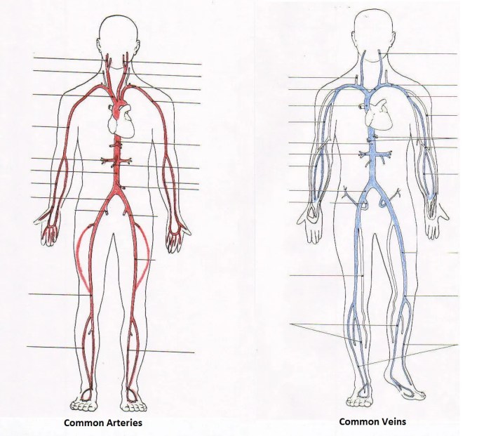
The veins of the trunk drain blood from the head, neck, and upper limbs into the heart. They are divided into two main groups: the superior vena cava and the inferior vena cava.
Superior Vena Cava
The superior vena cava is a large vein that drains blood from the head, neck, and upper limbs. It is formed by the union of the right and left brachiocephalic veins. The brachiocephalic veins are formed by the union of the internal jugular vein and the subclavian vein on each side.
The superior vena cava ascends through the mediastinum and enters the right atrium of the heart. It receives tributaries from the azygos vein, the hemiazygos vein, and the pericardial veins.
Azygos and Hemiazygos Veins
The azygos vein is a large vein that drains blood from the posterior chest wall and abdomen. It begins in the abdomen as the ascending lumbar vein and ascends through the thorax, passing behind the aorta. It receives tributaries from the intercostal veins and the bronchial veins.
The hemiazygos vein is a smaller vein that drains blood from the left side of the posterior chest wall. It begins as the ascending lumbar vein on the left side and ascends through the thorax, passing behind the aorta. It receives tributaries from the intercostal veins.
Inferior Vena Cava
The inferior vena cava is a large vein that drains blood from the lower limbs, abdomen, and pelvis. It is formed by the union of the right and left common iliac veins. The common iliac veins are formed by the union of the internal iliac vein and the external iliac vein on each side.
The inferior vena cava ascends through the abdomen and thorax, passing behind the liver and the heart. It receives tributaries from the renal veins, the hepatic veins, and the suprarenal veins.
Applied Anatomy
The clinical significance of the jugular venous pulse lies in its ability to provide information about the cardiac cycle and hemodynamics. The jugular venous pulse is a waveform that represents the pressure changes in the right atrium and jugular veins during the cardiac cycle.
It can be used to assess the heart rate, rhythm, and volume status, as well as to detect abnormalities in cardiac function, such as heart failure, valvular disease, and pericardial disease.
Cannulation of the Internal Jugular Vein
Cannulation of the internal jugular vein is a common procedure performed to obtain central venous access for the administration of fluids, medications, and blood products, as well as for monitoring hemodynamics. The technique involves inserting a catheter into the internal jugular vein, which is located in the neck.
There are two main approaches to cannulating the internal jugular vein: the anterior approach and the posterior approach.
- Anterior approach:In this approach, the needle is inserted just below the clavicle, at the level of the cricoid cartilage. The needle is then directed superiorly and medially towards the ipsilateral internal jugular vein.
- Posterior approach:In this approach, the needle is inserted at the level of the mastoid process, just behind the sternocleidomastoid muscle. The needle is then directed anteriorly and medially towards the ipsilateral internal jugular vein.
Potential Complications Associated with Central Venous Catheterization
Central venous catheterization is a relatively safe procedure, but it is not without potential complications. These complications can be divided into two categories: mechanical complications and infectious complications.
- Mechanical complications:These complications include pneumothorax, hemothorax, arterial puncture, and catheter-related thrombosis. Pneumothorax is the most common mechanical complication, occurring in approximately 1-2% of cases. Hemothorax is a rare but potentially life-threatening complication, occurring in less than 1% of cases.
- Infectious complications:These complications include catheter-related bloodstream infections (CRBSIs) and exit site infections. CRBSIs are the most common infectious complication, occurring in approximately 2-5% of cases. Exit site infections are less common, occurring in approximately 1-2% of cases.
Imaging Techniques
Imaging techniques play a vital role in the visualization, diagnosis, and management of venous disorders of the head and trunk. Various imaging modalities are employed, each with its unique advantages and limitations.
Ultrasound
Ultrasound is a widely used imaging technique that utilizes high-frequency sound waves to generate real-time images of the veins. It is non-invasive, portable, and relatively inexpensive.
Advantages:
- Real-time visualization of blood flow
- Assessment of vein patency, diameter, and flow velocity
- Detection of clots, stenosis, and other abnormalities
Disadvantages:
- Operator-dependent
- Limited penetration depth, especially in obese patients
- Difficult to visualize veins in certain anatomical locations
Computed Tomography (CT) Venography
CT venography involves injecting a contrast agent into the veins and obtaining cross-sectional images using X-rays. It provides detailed anatomical information and can detect abnormalities in both large and small veins.
Advantages:
- High spatial resolution
- Visualization of veins throughout the head and trunk
- Detection of deep vein thrombosis, pulmonary embolism, and other venous disorders
Disadvantages:
- Involves radiation exposure
- May not be suitable for patients with contrast allergies
- Can be expensive and time-consuming
Magnetic Resonance Imaging (MRI) Venography
MRI venography utilizes magnetic fields and radio waves to generate images of the veins. It does not involve radiation exposure and provides excellent soft tissue contrast.
Advantages:
- Non-invasive and radiation-free
- High soft tissue contrast
- Visualization of both large and small veins
Disadvantages:
- Expensive and time-consuming
- May require contrast agents
- Can be challenging for patients with claustrophobia or metal implants
Magnetic Resonance Angiography (MRA), Label the veins of the head and trunk
MRA is a non-invasive imaging technique that utilizes MRI technology to visualize blood vessels, including veins. It is similar to MRI venography but does not require contrast agents.
Advantages:
- Non-invasive and radiation-free
- Visualization of both arteries and veins
- Suitable for patients with contrast allergies
Disadvantages:
- Lower spatial resolution than CT venography
- May not be suitable for patients with claustrophobia or metal implants
- Can be time-consuming
Role of Imaging in Venous Disorders
Imaging techniques play a crucial role in the diagnosis and management of venous disorders of the head and trunk. They help identify the underlying cause of symptoms, such as pain, swelling, or thrombosis. Imaging can also guide treatment decisions, such as surgical or endovascular interventions.
By providing detailed anatomical information, imaging techniques facilitate the accurate diagnosis and appropriate management of venous disorders, leading to improved patient outcomes.
Questions Often Asked: Label The Veins Of The Head And Trunk
What is the largest vein in the body?
The superior vena cava
What is the function of the jugular veins?
To drain blood from the head and neck
What are the potential complications of central venous catheterization?
Infection, bleeding, and thrombosis

